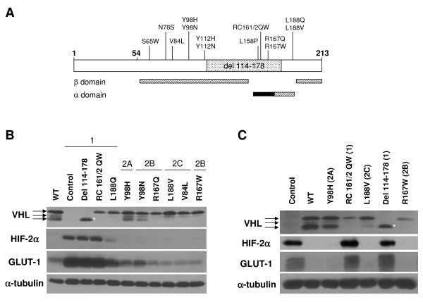
Figure 1.
Type 1, but not type 2 VHL mutants are grossly deficient in HIF-2α regulation. (A) Diagrammatic representation of pVHL, with first and last amino acid residues in bold. Codon 54 represents the start of the pVHL19 product. Mutations used in this study and location of the α and β domains in the reported VHL structure [84] are shown. The location of the elongin binding domain is demarcated with a black box. (B) RCC10 VHL-null cells were stably infected with retroviruses produced with the empty vector with no VHL insert (control) or with wild-type (WT) or mutant pVHL30 expression constructs, as indicated. Cells were grown to confluence and prepared cell lysates were equally loaded and separated by SDS-PAGE. Western blots were performed for VHL, HIF-2α, and GLUT-1. Note that the 3 bands observed for VHL are closely-migrating isoforms of pVHL30 [11], but are not pVHL19, which was not produced by these constructs (data not shown). α-tubulin was also assayed to demonstrate equal loading. *indicates decreased size of VHL deletion protein. (C) A498 VHL-null cells were similarly stably infected and the resulting cell lines were analyzed by western blotting.

