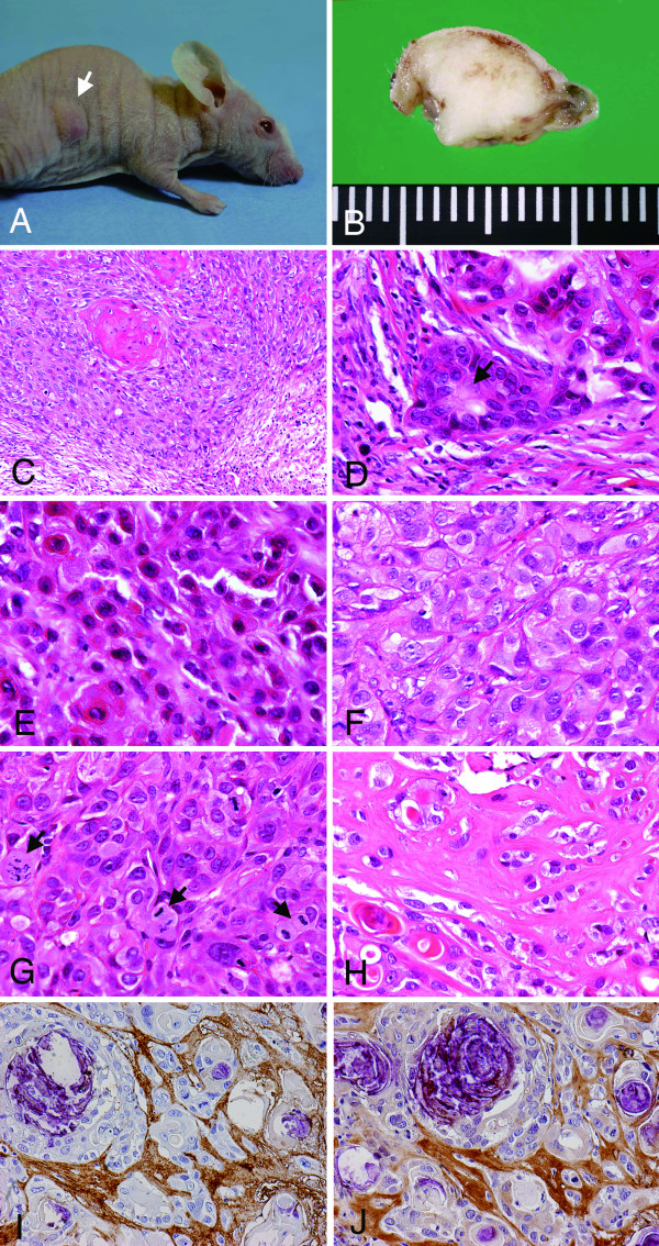
Figure 6.
Transplanted tumors of SM-AP cell systems in nude mice. Macroscopic view of a tumor mass by SM-AP5 in lateral back in a nude mouse (A); cut surface view of a subcutaneous tumor by SM-AP1 (B); histopathology of transplanted tumors by SM-AP4 (C), SM-AP1 (D, E), SM-AP5 (F, G), and SM-AP3 (H). HE stain, C, × 100; D-G, × 320; H, × 240; immunoperoxidase stains of SM-AP3 transplants for perlecan (I) and fibronectin (J), × 200, hematoxylin counterstain. SM-AP cells formed subcutaneous tumors measuring about 10 mm in diameter in nude mice within one to four months (A). The tumors were rather limited to the dermis expanding into the superficial part of the muscle layer but had no capsular structure (B). Histopathologically, the tumors were basically squamous cell carcinomas with definite tendencies towards keratinization with invasive natures, although there was no basal cell alignment along the periphery of the tumor cell nests (C). Around the tumor cell nests, myxoid stroma was induced. SM-AP1 to SM-AP3 cells formed mimics of ductal structures (D), and at the same time, SM-AP1 and SM-AP2 showed plasmacytoid appearances (E). SM-AP4 and SM-AP5 cells formed less differentiated carcinomas composed of tumor cells with ground-glass-like cytoplasm (F). Irrespective of tumors, mitotic figures were frequently observed (G), and the stromata were wide, hyaline, and poor in vascularity and lymphocytic infiltration (H). The hyaline stroma was immunopositive for perlecan (I) and fibronectin (J).

