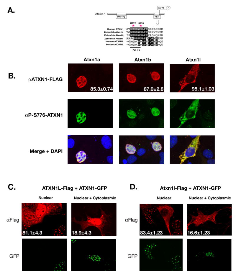
Figure 3. Zebrafish Atxn1/Axh proteins localize to the nucleus and are phosphorylated in tissue culture cells.

(A).ClustalW alignment was performed to show conservation of both the nuclear localization sequence and S776 residue among the zebrafish Atxn1/Axh proteins and human ATXN1 but not with mouse or human ATXN1L. (B). COS-1 cells were transfected with Flag-tagged zebrafish atxn1a, atxn1b, or atxn1l. Twenty-four hours post transfection these cells were stained using an α-FLAG or α-P-S776 ATXN1 antibody. Zebrafish Atxn1a and Atxn1b localized to the nucleus of the cell while Atxn1l was mostly cytoplasmic. All three proteins were recognized by the phospho-specific ATXN1 antibody. (Each transfection was done in triplicate and 400 cells per experiment were scored for protein localization. The percent of cells with nuclear (Atxn1a and Atxn1b) or cytoplasmic (Atxn1l) localization is noted in the αFlag panel). (C). Flag-tagged human ATXN1L was cotransfected with GFP-tagged human ATXN1 into COS-1 cells and stained with an α-FLAG antibody. Human ATXN1L alone localized to both the nucleus and cytoplasm of the cell (as seen by the two cells not transfected with ATXN1 in the nuclear panel) but when coexpressed with ATXN1, became predominantly nuclear. In a small percentage of cells, cytoplasmic and nuclear localization of ATXN1L was observed in the presence of ATXN1. (This transfection was performed in triplicate, and 100 cotransfected cells per experiment were scored for nuclear or nuclear and cytoplasmic localization of ATXN1L. The percentages are indicated in each representative image.) (D). Flag-tagged zebrafish atxn1l was cotransfected into COS-1 cells with GFP-tagged human ATXN1. When expressed with human ATXN1, zebrafish Atxn1l became predominantly nuclear in contrast to the mostly cytoplasmic location of zebrafish Atxn1l when expressed alone (A). In a small percentage of cells, zebrafish Atxn1l was visualized in both the cytoplasm and nucleus in the presence of human ATXN1. (This transfection was performed in triplicate and 100 cotransfected cells per experiment were scored for Atxn1l localization. Percentage nuclear or nuclear and cytoplasmic is noted in each representative image.)
