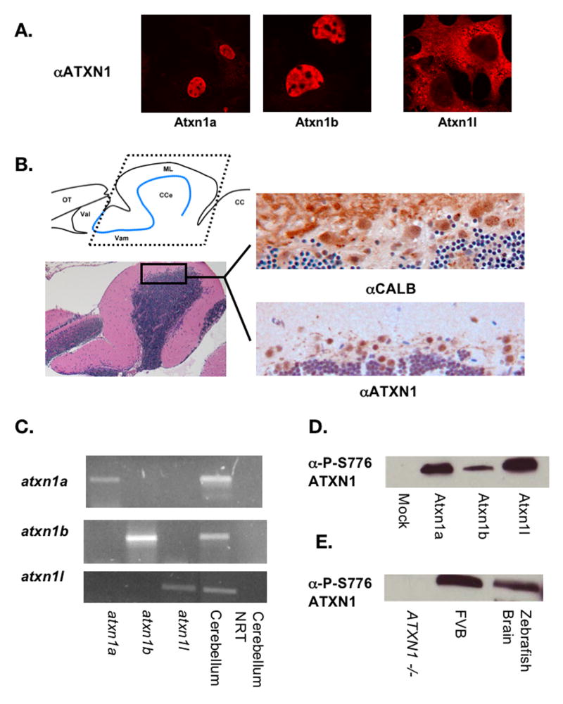
Figure 4. Zebrafish Atxn1/Axh proteins are expressed and phosphorylated in the adult cerebellum.
To demonstrate that the anti-ATXN1 antibody used in these studies cross-reacts with all three zebrafish Atxn1/Axh proteins, COS-1 cells were transfected with Flag-tagged zebrafish atxn1a, atxn1b, or atxn1l. Twenty-four hours post transfection, the cells were stained with the anti-ATXN1 antibody (A). Immunohistochemistry was performed on sections from paraffin embedded zebrafish heads using the anti-ATXN1 antibody tested in panel A and an anti-calbindin antibody as a Purkinje cell marker (B). Expression of the zebrafish Atxn1 proteins was detected in the cerebellar Purkinje cells. Both a diagram of the zebrafish cerebellum (with the Purkinje cell layer highlighted in blue) as well as an H&E section are included to provide an orientation to the zebrafish cerebellum. To determine which ATXN1 homologs are expressed in the cerebellum, RT-PCR analysis was performed on RNA isolated from adult zebrafish cerebellum to detect expression of zebrafish atxn1a, atxn1b, or atxn1l (C). RNA from COS-1 cells transfected with each construct individually was used as a control. To determine if the zebrafish Atxn1/Axh protein family is phosphorylated at S776 in vivo, western blot analysis was performed using an anti-P-S776 ATXN1 antibody. Protein lysates from COS-1 cells transfected with zebrafish atxn1a, atxn1b, or atxn1l (D) and lysates from a zebrafish brain (E) were probed with an anti-P-S776 antibody. Mock transfected cells (D) and wild type (FVB) and ATXN1 -/- mouse brains (E) were included as controls. (OT = Optic Tectum, VaL = Lateral Valvula Cerebelli, VaM = Medial Valvula Cerebelli, CCe = Corpus Cerebelli, ML = Molecular Layer, and CC = Crista Cerebelli)

