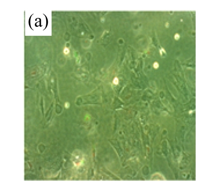
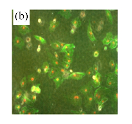
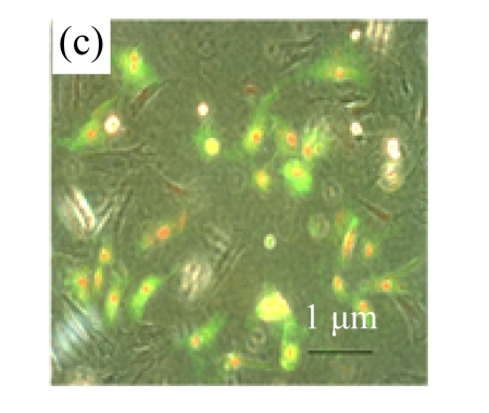
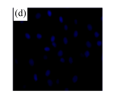
Fig.2.
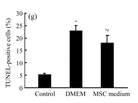
MSC medium reduced H/R-induced apoptosis of cardiomyocytes
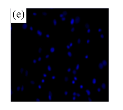
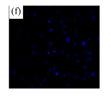
(a)~(c) Apoptotic cells were detected by Annexin V-FITC staining for labeling early-stage apoptotic cells (green) and necrotic cells (PI stained, red); (d)~(f) Hoechst 33342 staining of cardiomyocytes: apoptotic cells were characterized by nuclear shrinkage with condensed chromatin structure; (g) Quantification of apoptotic cardiomyocytes measured by TUNEL assay. The fraction of apoptotic cells was determined in five random microscopic fields totalling at least 1000 cells/group. Cardiomyocytes were hypoxic for 24 h and were reoxygenated for 3 h in serum-free DMEM (DMEM group) or the medium abstracted from MSCs (MSC medium group). Control cells were cultured in DMEM containing 20% (w/v) fetal calf serum. Results are representative of three independent experiments. Data are shown as mean±SEM. Control group: (a), (d); DMEM group: (b), (e); MSC medium group: (c), (f). * P<0.05 vs control group, # P<0.05 vs DMEM group

