Fig.3.
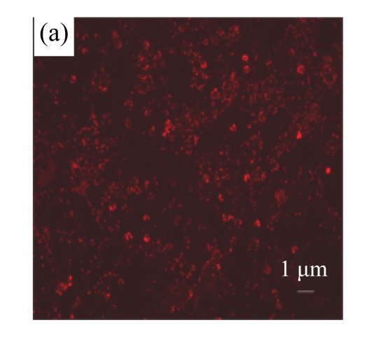
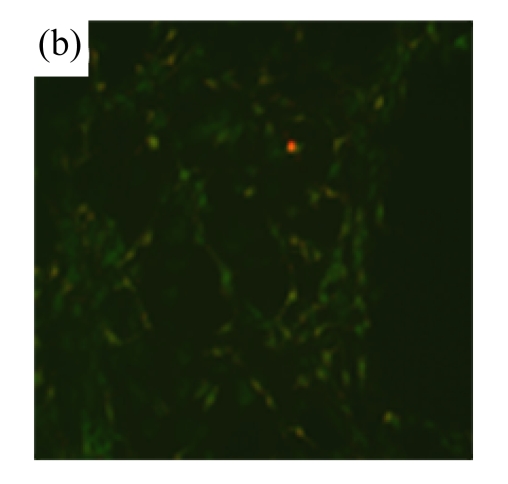
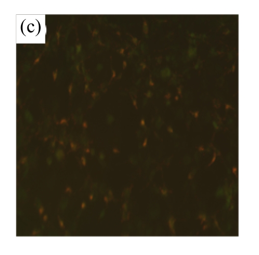
MSC medium attenuated the reduction of mitochondrial membrane potential of H/R-induced cardiomyocytes
Mitochondrial membrane potential was determined using the potential-sensitive fluorescent probe JC-1. (a) Normally cultured cardiomyocytes contained red fluorescent mitochondria in the cytoplasm; (b) Cardiomyocytes after treatment with H/R and serum-free DMEM culture showed green fluorescence, indicating the loss of mitochondrial membrane potential; (c) Cardiomyocytes with H/R and MSC medium culture showed red fluorescent mitochondria in the cytoplasm, indicating the preservation of the mitochondrial membrane potential. Results are representative of three independent experiments


