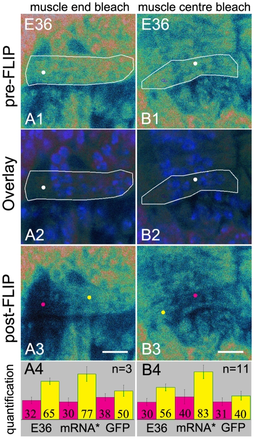
Figure 5. Endogenous RNAs are also limited with respect to their movement in myofibers.
FLIP experiments in the muscle 12 of wild type Drosophila after injection of Hoechst and E36. (A1, B1) Pre-FLIP confocal images. (A2, B2) Overlays of pre-FLIP images and confocal images showing Hoechst stained nuclei confirming that the bleachspots are located outside the nuclei (white dots). (A3, B3) Post-FLIP confocal images. Bars, 8 µm. (A4, B4) Quantification. Information on white lines, white, yellow and pink dots and the bar graphs are given in the legend of Fig. 2. (A3) Bleaching at the muscle end. Fluorescence is lost from the muscle end but remains present in the rest of the cell. (B3) Bleaching at the muscle centre. Fluorescence is lost from the muscle centre and remains present at the both muscle ends. These data are compared with the FLIP data from GFP and the reporter mRNA expressing myofibers (Fig. 2) (A4, B4) Bar graphs show that the fluorescence in the bleached spots (pink) is reduced and that the fluorescence outside the bleached domains (yellow) is also diminished but to a lesser extent than when FLIP experiments were performed in GFP expressing myofibers. mRNA* = GFP-tagged mRNA. For quantification of the fluorescence see Materials and Methods.

