Figure 2.
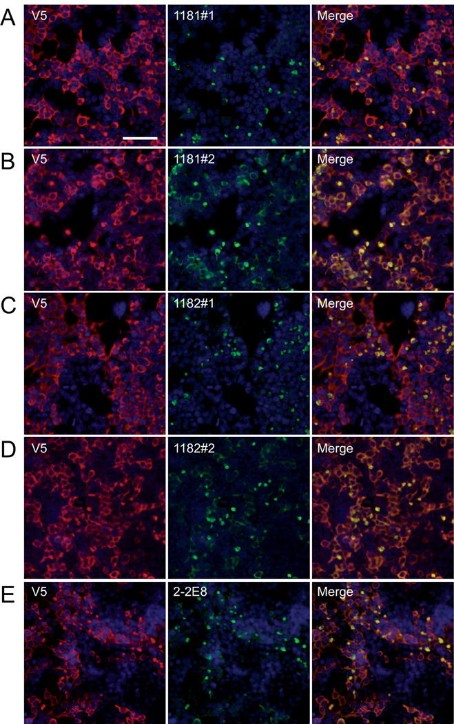
Immunofluorescence of leucine-rich repeat kinase-2 (LRRK2) in transfected 293t cells. Double immunofluorescence was performed on 293t cells transfected with LRRK2-V5, and immunoreactivity was compared between monoclonal anti-V5 (red, A–D) or polyclonal anti- V5 (red, E) and LRRK2-specific antibodies (green) 1181#1 (A), 1181#2 (B), 1182#1 (C), 1182#2 (D) or 2-2E8 (E). Staining with anti-V5 antibody, LRRK2 appeared mostly cytosolic. Some LRRK2-specific antibodies preferentially recognized aggregated forms of LRRK2, observed as perinuclear aggregates in a subpopulation of cells. 1181#1 (A) and 1182#1 (C) showed greater specificity for the aggregated form, while 1181#2 (B) and 1182#2 (D) recognized aggregated and cytosolic forms of LRRK2. The monoclonal LRRK2 antibody 2-2E8 (E) also specifically recognized these aggregates. Bar scale: 100 µm.

