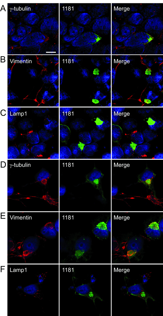
Figure 6.
Confocal microscopy showing colocalization of LRRK2 with markers of aggresome formation. (A–C) 293t cells were transfected with LRRK2, and (D–F) COS-7 cells were transfected with LRRK2 and treated with MG132 (10 µM, 16 hours) and then double immunofluorescence was performed between aggresome markers anti-γ-tubulin (A, D), antivimentin (B, E), or anti-lamp1 (C, F) and 1181#1. Colocalization was observed for both γ-tubulin and vimentin. Some lamp1 immunofluorescence was noted around the LRRK2 aggregate. All images provided are of a single Z-plane of <0.7 µm. Bar scale: 10 µm.

