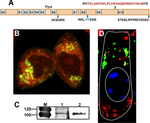
Fig. 7.
Expression of mitochondrial BKα in vitro and in vivo. A, splice sequences shown in relation to the different regions of a BK-DEC variant cloned from mouse cochlea and used in CHO expression studies. B, live CHO cells transfected with pcDNA3.1 containing Cerulean in fusion with the C terminus of the BK-DEC variant and treated with MitoTracker (red) to identify mitochondria. Cerulean was pseudo-colored (dark green) to visualize overlap with red as yellow. Dark green immunostaining of BKα alone is observed at the plasmalemma. C, immunoprecipitation of BKα, using a pure mitochondrial preparation from mouse cochlea (left lane) and brain (right lane), shows bands at the expected weight of BK. D, tangential section of an outer hair cell, as outlined in white, showing the supranuclear region where BKα (red) is colocalized (yellow) in mitochondria with VDAC channels (green). The nucleus (oval) is partially stained with blue DAPI.

