
Figure 7.
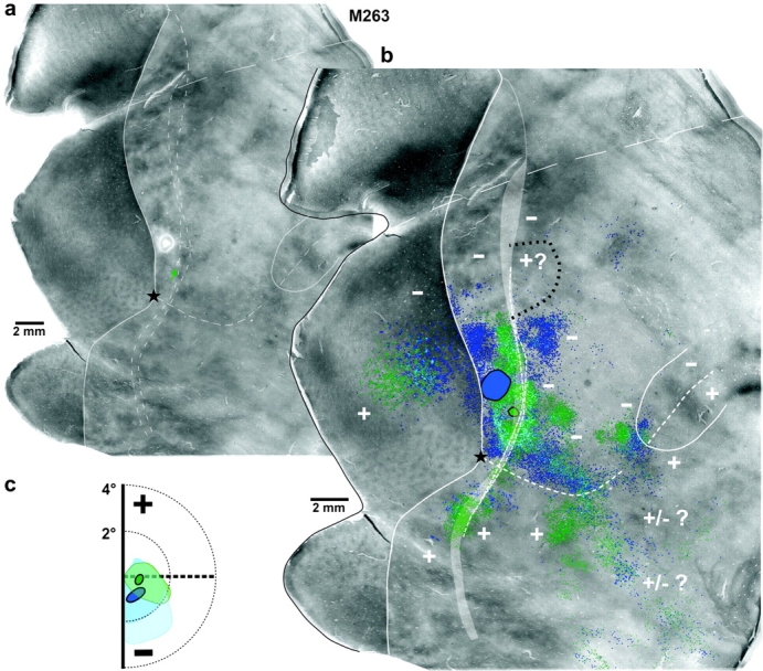
Two additional examples of foveal lateral injection cases. Case M263. (a) CO image. The green arrow points at the small CO-pale spot in V2d corresponding to the CTB488 injection site. The much larger CO-pale spot just above it is the FB injection site. (b) The same CO image is shown enlarged with overlaid 2 composite injection sites (encircled in black) and the mapped label (CTB488—green, FB—blue) resulting from them. Light blue shows regions of overlap of the 2 different labels. (c) Visual field map of the CTB488 (green circle) and FB (blue oval) injection sites and resulting intra-areal label in V2d and V2v (pale green and blue, respectively; lighter blue indicates less dense FB label). Other conventions are as in Figure 4.