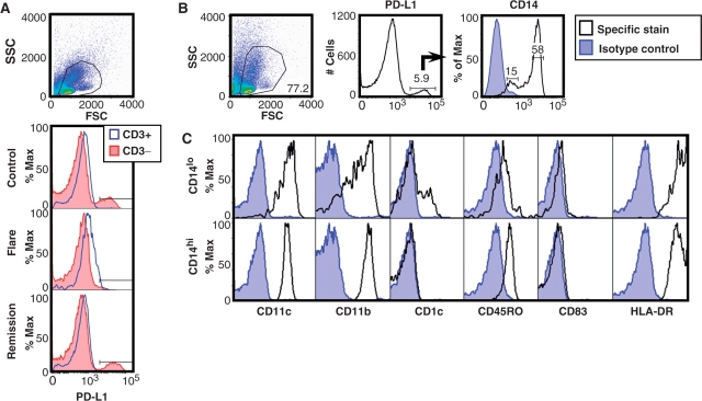
Fig. 1.
Primary human PBMC spontaneously up-regulate PD-L1 protein. (A) Cells were cultured for 1 day and live PBMC gated by forward and side scatter. Histograms show mean fluorescence intensity (MFI) of PD-L1 as measured on CD3+ and CD3− cells from a healthy paediatric control (top), from a patient in SLE flare (middle) and from the same patient during SLE remission (bottom). (B) Using control PBMC, PD-L1+ cells were further characterized and found to separate into two groups based on CD14 expression. (C) Using antibodies to phenotypic markers, the PD-L1+ CD14lo and CD14hi cells were identified as immature mDC and Mo, respectively.

