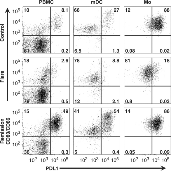
Fig. 3.

Lupus flare APC express positive co-stimulatory molecules. PBMC were cultured for 1 day and gated for APC as given earlier. Compared with control cells (top row), SLE flare APC (middle row) lacked PD-L1 and the CD80/86hi subset of mDC. In contrast, SLE remission cells (bottom row) expressed both PD-L1 and CD80/86 in a pattern similar to that of controls (numbers in each graph represent the percentage of cells in each quadrant). Results are representative of two separate experiments using PBMC from three healthy controls, two SLE flare and two SLE remission.
