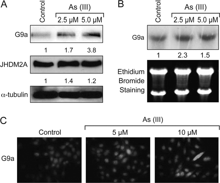
Fig. 3.
Arsenite-induced histone methyltransferase G9a expression. A549 cells were treated with arsenite (2.5 and 5 μM) for 24 h. A set of representative results is shown from three independent experiments. (A) G9a and JHDM2A protein levels were analyzed by western blotting with antibodies as described in the Materials and Methods. The same membrane was reblotted with α-tubulin to assess protein loading. (B) Arsenite exposure increases G9a mRNA in A549 cells. Total RNA was extracted and subjected to northern blotting. The bottom panel shows the total RNA in the formaldehyde–agarose gels detected by ethidium bromide staining. The numbers below the figure represent the relative intensity of the bands. (C) Increased level of G9a in As-treated A549 cells. A549 cells were treated with arsenite at different concentrations for 24 h, followed by immunofluorescent staining with G9a antibody as described in Materials and Methods.

