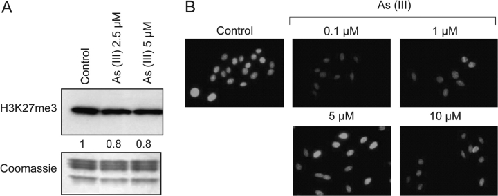
Fig. 5.
Decreased level of H3K27 trimethylation in arsenite-treated A549 cells. A549 cells were treated with arsenite at different concentrations for 24 h, and trimethylated H3K27 was detected by western blotting (A) and immunofluorescent staining (B). The western blot result was repeated three times and representative results are shown and quantitated. Coomassie blue was used to assess equal histone loading. The numbers below the figure represent the relative intensity of the bands.

