
Figure 2. A beating heart is required for HSC formation and artery development.
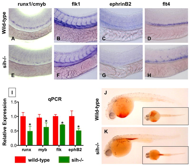
(A-H) Effect of sih mutation on HSC and vascular formation at 36hpf.
(A,E) runx1/cmyb expression is greatly reduced in sih-/- embryos compared to WT siblings.
(B,F) flk1 expression reveals a grossly normal vascular pattern in sih-/- embryos; changes in the development of the intersomitic vessels and vascular plexus were noted in some animals.
(C,F) ephB2 expression is diminished in sih-/- embryos.
(D,H) flt4 expression is expanded in sih-/- embryos.
(L) The expression levels of HSC (runx1, cmyb), vascular (flk) and arterial (ephB2) markers are significantly decreased in sih-/- embryos compared to sibling controls (t-test, p<0.05, n=3), as measured by qPCR at 36hpf.
(J,K) The sih mutation has no effect on primitive hematopoiesis as seen by benzidine staining at 36hpf; in the absence of a heartbeat blood is pooled in the major vessels.