
Figure 5. nos1 is required for HSC formation in zebrafish.
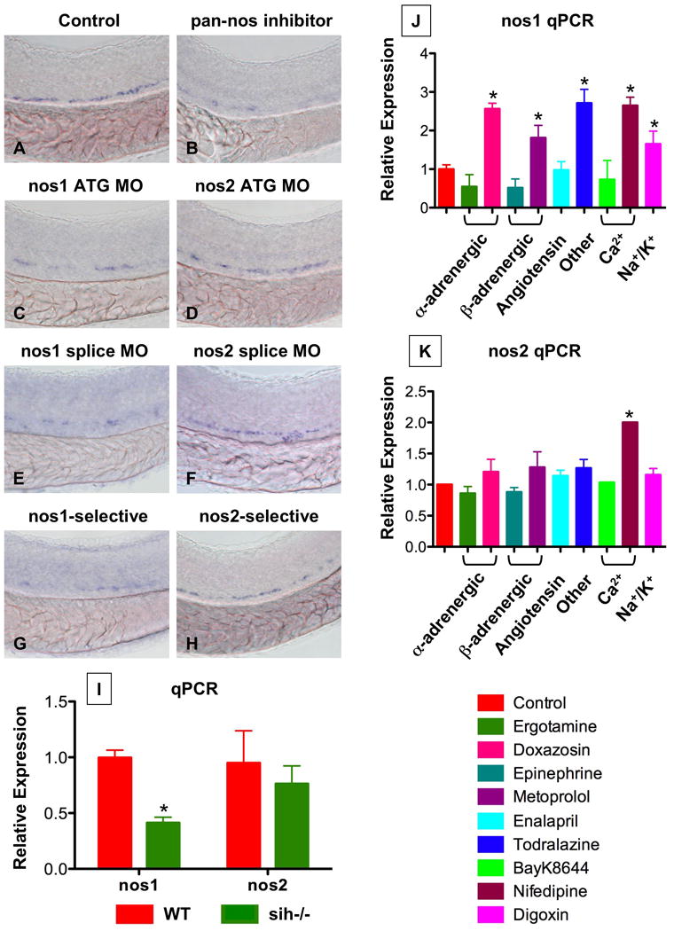
(A-H) In situ hybridization for runx1/cmyb at 36hpf.
(C,E) nos1 knockdown (40 μM) decreased HSC formation.
(D,F) MO (ATG and splice site) against nos2 (40 μM) had no effect on HSC development.
(B,G,H) Chemical nos inhibition confirmed the specific requirement for nos1: Embryos exposed to non-specific (L-NAME; 10 μM) and nos1-selective (S-methyl-L-thiocitrulline; 10 μM) inhibitors demonstrated decreased HSC formation; nos2-selective inhibition (1400W; 10 μM) had minimal impact.
(I) WT and sih-/- embryo extracts (n=20) were subjected to qPCR (* nos1; WT vs. sih, t-test, p<0.001, n=3; nos2; WT vs. sih, p=0.385, n=3).
(J,K) Effect of flow modifying chemicals (10 μM, 10 somites-36hpf) on nos1 and nos2 expression; nos1 is significantly regulated by most compounds tested. * significant vs. control, ANOVA, p<0.01, n=3.