
Figure 6. The effect of NO signaling on HSC development is cell-autonomous.
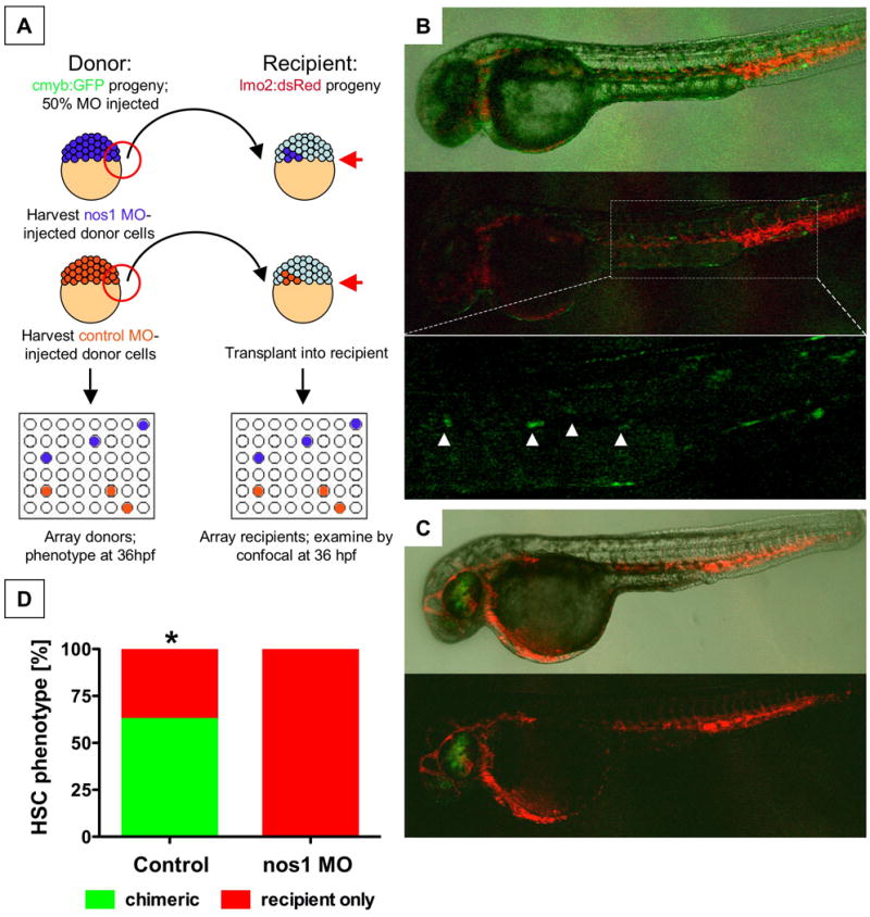
(A) Cells from cmyb:GFP transgenic donor embryos, injected with nos1 ATG MO or control MO, were transplanted into lmo2:dsRed recipients at the blastula stage.
(B) Donor contribution to HSC formation assessed by confocal microscopy at 36hpf. Shown are the merged picture on the top, red/green merge in the middle, and a high-magnification view of green fluorescence only on the bottom. cmyb:GFP donor-derived HSCs in recipients are highlighted by arrowheads.
(C) nos1 MO donors never contributed to HSC formation; presence of cmyb:GFP-derived donor cells in the eye is indicative of a successful transplant.
(D) HSC chimerism in transplanted embryos (control vs. nos1 MO, Fisher's exact, p=0.0065, n≥8).