Figure 4.
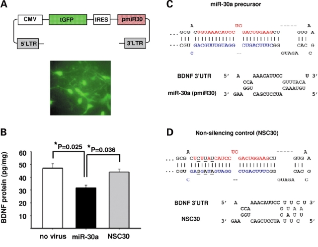
MiR-30a mediates translational inhibition of BDNF in neurons. Neuronal cultures from rat forebrain were infected with lentiviruses that contained constructs co-expressing GFP and precursor miRNAs. (A) Top: Map of the lentiviral vector used; Bottom: Representative image of neuronal culture infected with the GFP-expressing lentiviral vector shown above. (B) Bar graphs showing BDNF protein levels (mean ± SEM) for cultures infected with miR-30a (n = 3), or miR-NSC30 (n = 3) (see text for details), and non-infected (‘no virus’, n = 2) cultures. Notice the significant decrease in BDNF protein in miR-30a overexpressing cultures. P-values after post-hoc Tukey/ANOVA. (C and D) Top: Secondary structures of (C) miR-30a precursor and (D) miR-NSC30 which contains a 3 base substitution in the seed sequence of miR-30a-5p. Mature miR-30a-5p is depicted in red, miR-30a-3p in blue and the nucleotide changes in the non-silencing control, NSC30, are shown in black and underlined. Bottom: Predicted interactions between the first target site in BDNF 3′-UTR (see text for details) and either (C) wild-type miR-30a-5p or (D) non-silencing precursor (‘NSC30’).

