
Figure 3.
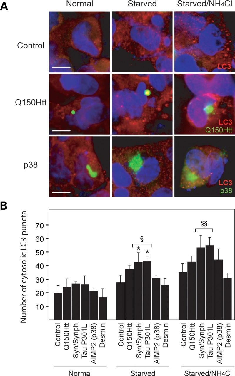
Formation of AIMP2 (p38)- and desmin-positive inclusions does not impair the autophagic system. (A) Representative confocal images showing LC3 staining in untransfected SH-SY5Y cells (control) (top panels), or cells containing either Q150Htt-positive inclusions (middle panels) or AIMP2 (p38)-positive inclusions (bottom panels) cultured under different conditions (as indicated) following proteasome inhibition. (B) Bar graph showing the average number of cytosolic LC3 puncta per cell in control cells or those containing various types of inclusions (as indicated) cultured under different conditions following proteasome inhibition. Each of these experiments was repeated at least three times (*P < 0.05 versus respective value under normal conditions, §P < 0.01, §§P < 0.001 versus control cells under same conditions).