
Figure 7.
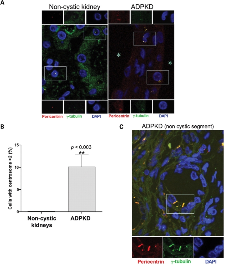
Centrosome amplification occurs in ADPKD patients. (A) Pericentrin (red) and γ-tubulin (green) immunostaining of DAPI-counterstained archival tissues from control non-cystic and ADPKD human kidneys (original magnification ×1000). The large frames show the merged fields while individual staining of centrosomal markers corresponding to the near insets are further magnified at the top and the bottom. Shown are representative images from one of five non-cystic controls and one of three ADPKD patients. (B) Scores of cells with more than two centrosomes were obtained from different non-cystic and ADPKD patients (each group n = 3; total count of randomly chosen cells >600). Statistical analysis was performed using the unpaired two-tailed t-test with Welch correction. (C) Centrosome amplification was observed also in epithelial cells within non-cystic tubules from ADPKD specimens. The asterisks mark the lumen of the cysts.