Figure 2.
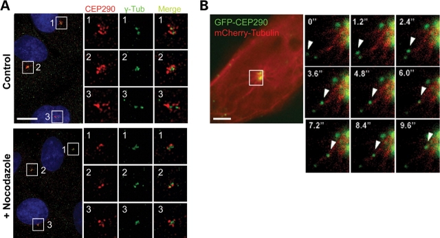
CEP290 moves along microtubules to the centrosome. (A) Cultured hTERT-RPE cells were treated with 20 µm nocodazole for 2 h and double-stained with anti-CEP290 and anti-γ-tubulin antibodies. Nocodazole treatment causes substantial decrease in granular CEP290 staining detected around the centrosome, suggesting that the recruitment of CEP290 to centriolar satellites requires intact microtubules. (B) Time-lapse observation of the movement of GFP-CEP290 along mCherry-labeled microtubules in live IMCD-3 cells. The boxed area in the left panel is magnified in the small panels. Numbers indicate the time lapse in seconds. Scale bars: (A) 10 µm; (B) 5 µm.

