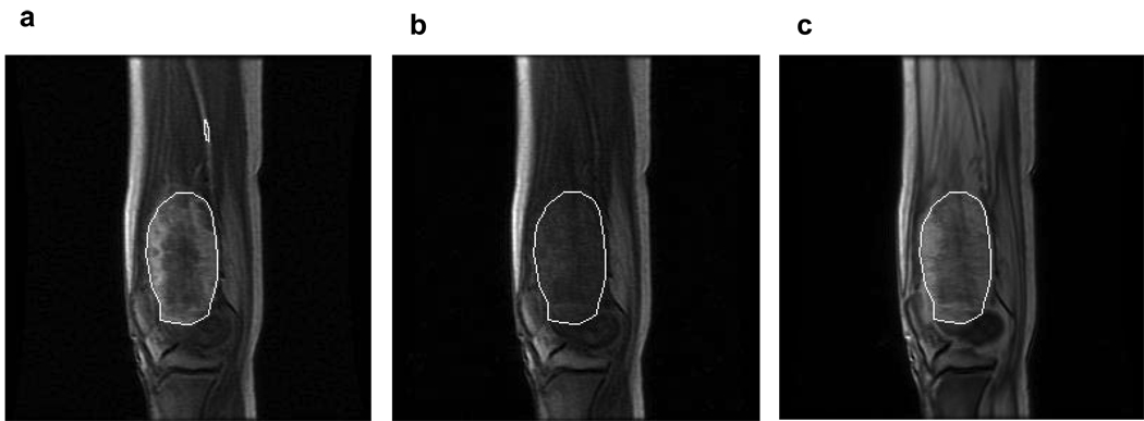
Figure 1.
Sagittal images from a patient with an osteosarcoma in the distal femur: (a) A post-contrast image extracted from a multi-slice dynamic contrast-enhanced (DCE) MRI acquisition, with the white ROI circumscribing the contrast-enhanced tumor. The yellow ROI was placed within the adjacent femoral artery for arterial input function (AIF) data sampling. (b) The pre-contrast image from the same DCE-MRI series with the same location as in panel a. (c) A proton density image acquired prior to DCE-MRI with the same location and slice thickness as panels a and b. The white ROIs in panels b and c were positioned in the same spatial location as in panel a.

