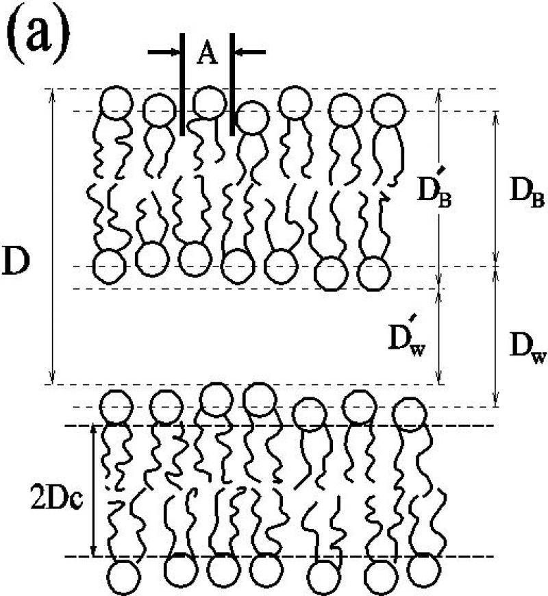
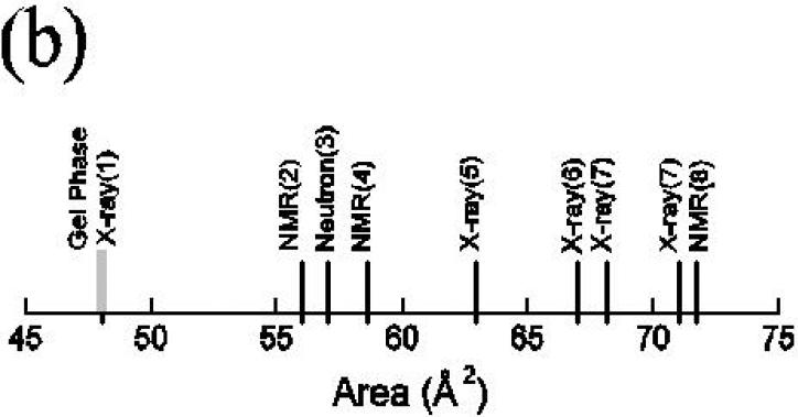
Fig. 1.
(a) The sketch of two bilayers in a multilamellar vesicle (MLV) identifies the primary lamellar repeat spacing D, the area A per molecule, the hydrophobic thickness 2DC the Luzzati thickness DB, the water thickness DW, the steric thickness , and the steric water thickness . (b) Prominent literature values for A for DPPC in the Lα phase (black) compared to the gel phase (grey).


