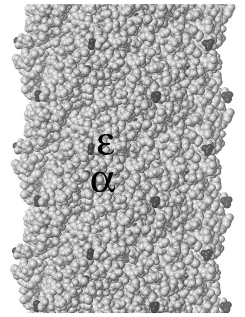
Figure 1.
Space-filling model (including hydrogens) of a short section of the tubular sheath of filamentous bacteriophage fd (Protein Data Bank accession no. 2C0X, pdb.org; see References 4 and 5), with amino groups highlighted in black. The section depicted includes all or parts of 30 pVIII subunits (out of 2700 for wild-type virions, 4000 or more for some phage-display constructs), each with a highly exposed ε-amino group on the lysine at position 8 and a mostly buried α-amino group on the N-terminal alanine; only some of the α-amino groups are partly visible in the image. The overall diameter of the sheath is ~6 nm.

