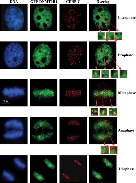
Figure 4.
A fraction of DNMT3B co-localizes with CENP-C particularly during metaphase. HeLa cells were transfected with GFP-tagged DNMT3B1 (green panels), synchronized with a double thymidine block, released, then fixed when cells were in mitosis. Interphase cells were also examined. Transfected cells were stained with anti-CENP-C (red panels) antibody. DNA was stained with DAPI (blue panels). An overlay of the red and green channels is shown in the right-most panels. Representative images of cells in interphase (top row) or the different phases of mitosis (lower four rows) are shown. Select regions are enlarged in the small red-boxed regions of the overlay panel to highlight closely opposed or overlapping DNMT3B and CENP-C foci. Double red arrowheads indicate closely juxtaposed foci. Scale bar—10 µm.

