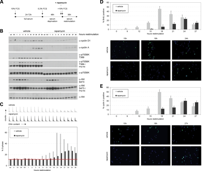
Figure 2.
Blocking mTORC1 activity delays S-phase induction without effects on mTORC2 activity. IMR-90 fibroblasts, synchronized in G0/G1 via serum deprivation, were serum restimulated to re-enter the cell cycle in the presence or absence of the mTOR inhibitor rapamycin. (A) Schematic outline of the synchronization procedure. Briefly, cells growing under full serum (10% FCS) conditions for 24–72 h were serum-deprived in medium containing 0.2% serum (0.2% FCS) for 48 h and serum restimulated (10% FCS) for additional 36 h. Rapamycin was added 30 min prior to restimulation at a concentration of 100 nm. Control cells were treated with an equal volume of DMSO and analysed in parallel. (B) Cells treated as described in (A) were lysed and examined for the expression level of cyclin D1, cyclin A, p70S6K T389, p70S6K, Akt S473 and Akt at the indicated time points via immunoblotting. (C) In addition, cells were cytofluorometrically analysed for DNA content at the indicated time points. DNA profiles are presented (upper panel). In addition, the percentage of cells in S-phase was determined for DMSO (vehicle)- and rapamycin-treated cells. To facilitate interpretation of S-phase induction, a baseline corresponding to the S-phase values of serum-deprived vehicle- and rapamycin-treated cells was inserted into the graph. (D) Experiments as described in (A, B and C) were repeated and the percentage of cells actively undergoing DNA replication was analysed by immunocytochemical BrdU incorporation assay at specific time points as indicated (at least 250 cells were scored for each timepoint). (E) In addition, cells derived from the same pool of cells as described in (D) were immunocytocemically stained for cyclin A expression (at least 250 cells were scored for each timepoint).

