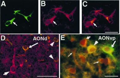
Figure 3.
Neurotransmitter expression by transplanted NRP cells. Sections from brains transplanted with GFP-NRP cells were double-labeled with anti-GFP along with anti-ChAT (A–C), antiglutamate (D), and anti-GABA (E). Anti-GFP is visualized with a FITC-conjugated secondary. (A–C) A representative section from the occipital cortex. Images from A and B are superimposed in C showing NRP cells uniformly expressing ChAT (arrow). (Bar = 100 μm.) (D) A representative confocal image from the AONd shows a host neuron expressing glutamate (short arrow) and transplanted NRP cells expressing GFP and glutamate (long arrow, arrowheads). (Bar = 100 μm.) (E) A representative section from the AONvp showing a GABA (−) NRP cell, (long arrow), GABA (+) host neurons (short arrow, thin arrows), and a GABA (+) GFP-NRP cell (arrowhead). (Bar = 50 μm.)

