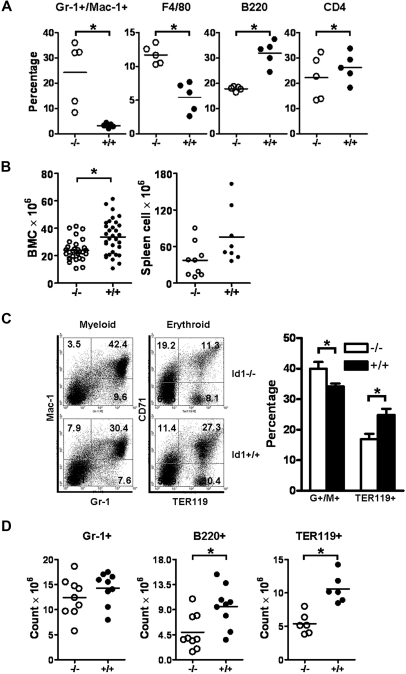
Figure 1.
Id1−/− mice show decreased BM cellularity with fewer myeloid, erythroid, and lymphoid cells but increased neutrophils and monocytes in the PB. (A) PBCs obtained from Id1−/− (n = 5) and Id1+/+ (n = 5) mice were analyzed with lineage-specific monoclonal antibodies. The horizontal bars indicate the mean percentage (*P < .01). Data were obtained from 2 independent experiments. (B) Total BM (Id1−/−, n = 30; Id1+/+, n = 32) and spleen (Id1−/−, n = 9; Id1+/+, n = 8) cells were enumerated by the use of a hemacytometer (*P < .01). (C) Left panel shows the flow cytometric analysis for granulocytes/monocytes (Gr-1 × Mac-1) and erythroid cells (TER119 × CD71) in Id1−/− and Id1+/+ BMC. The numbers in each quadrant indicate the percentage of each cell type among the total analyzed cells. Right panel shows percentages of Gr-1+/Mac-1+ or TER119+ BMC from all Id1−/− (n = 9) and Id1+/+ (n = 6) mice analyzed (*P = .01). (D) The absolute number of Gr-1+, B220+, and TER119+ BMC was calculated by multiplying the fraction of positive cells to total BMC (*P < .01). Data were obtained from 3 independent experiments, and the horizontal bars indicate the mean cell counts.

