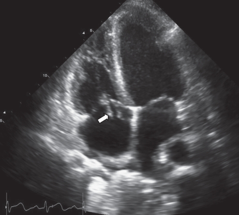
Figure 3).
Apical four-chamber view of an echocardiogram performed seven days after treatment, demonstrating significant improvement in the appearance of the vegetation since the previous echocardiogram. There is no longer any mass visible in the left atrium. There are two masses on the tricuspid valve, measuring 15 mm × 5 mm, and 13 mm × 4 mm (arrow indicates the larger mass)

