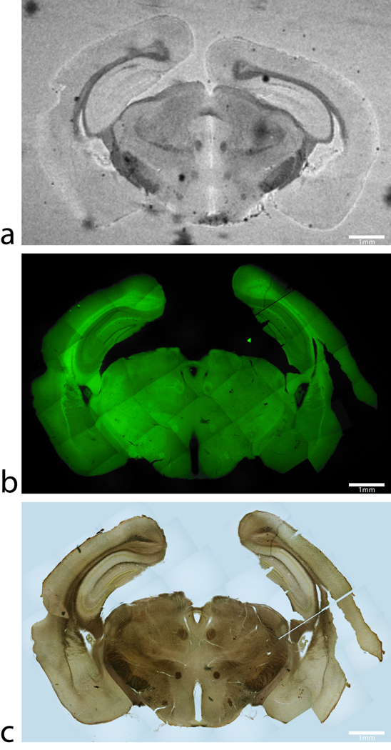
FIG 4.
Magnetic resonance image, thioflavin-S and iron stains from a control C57BL/6 mouse. (a) An MGE T2* weighted image, (b) thioflavin-S, and (c) Perl’s iron stain of same 60 µm thick section of tissue from a C57BL/6 control mouse at approximately −2.80mm Bregma. The T2* weighted image shows hypo-intensities and iron staining at regions of known high iron concentration such as the substantia nigra, white matter tracks and the caudate/putamen. Thioflavin-S staining reveals only non-specific background staining with no beta-amyloid plaques in the control animals. There are no MR hypo-intensities that are associated with positive thioflavin-S staining. Scale bars in all images are 1mm.

