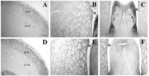
Figure 2.

Immunhistochemical analysis of MEF2A and MEF2D in the rat forebrain. MEF2A (A-C) and MEF2D (D-F) expression were analyzed in the cortex (A,D), striatum (B,E), and the septum (C,F) of the adult rat by immunoperoxidase staining. In the cortex MEF2A- (A) and MEF2D-ir (D) were seen in nuclei of neurons across all cortical lamina. MEF2A- (B) and MEF2D-ir (E) nuclei were also seen throughout the striatum. In contrast, although MEF2D-ir is localized to nuclei in the septum (F), extensive MEF2A-ir processes are present in the LS/BNST (C). (*, lateral ventricle, cortical layers are indicated by Roman numerals). Scale bar = 500 μm.
