Figure 3.

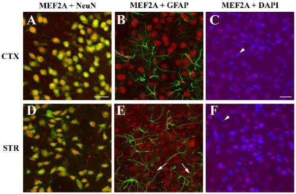
MEF2A is expressed in neurons but not astrocytes. Cell type specific expression of MEF2A was assessed in the cortex (A-C) and striatum (D-E). MEF2A-ir (red) was almost always localized to nuclei in the cortex and striatum, although rare MEF2A-ir fiber-like structures can be seen in the medial (periventricular) striatum (E, arrows). Double-labeling experiments with antibodies against MEF2A (red) and the neuronal marker NeuN (green) reveals that virtually all NeuN positive cells express MEF2A in the cortex (A) and the striatum (B). In contrast, MEF2A-ir (red) does not colocalize with astrocytes labeled with GFAP (green) in the cortex (B) or the striatum (E). Counter-staining cortical (C) or striatal (F) sections with the nuclear stain DAPI (blue) reveals that virtually all large nuclei characteristic of neurons are MEF2A-positive (red), whereas in the smaller nuclei, typically associated with glial cells, MEF2A-ir is absent (C,F arrow head). Scale bar = 25 μm in A also applies to B, D, E; scale bar = 25 μm in C also applies to F.
