
Figure 7.
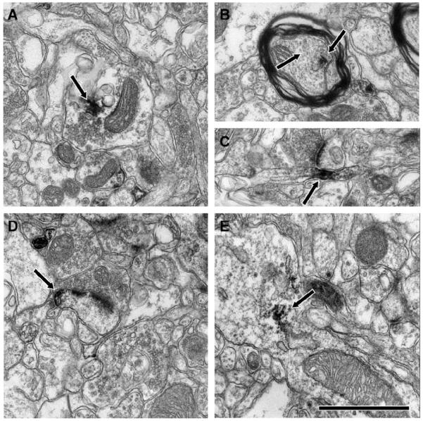
Subcellular localization of MEF2A-ir in the LS/BNST. Immunoelectron microscopic analysis revealed MEF2A-ir in both axons and dendrites. Labeled axon terminals were particularly common (A, arrow). Preterminal myelinated (B, arrow) and unmyelinated (C, arrow) axons also contained MEF2A-ir. In addition, MEF2A-ir dendritic spines (D, arrow) and shafts (E, arrow) can be observed. Scale bar = 500 nm.