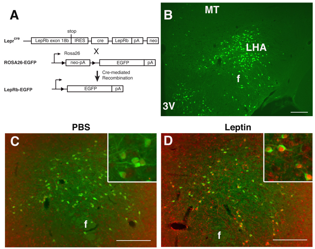
Figure 1. LepRbEGFP mice reveal a large population of leptin-responsive LHA LepRb neurons.
(A) Schematic diagram demonstrating cre-mediated EGFP expression in LepRb expressing cells of LepRbEGFP mice. (B) Immunofluorescent detection of EGFP (green) in the LHA of LepRbEGFP mice. (C,D) Immunohistochemical detection of pSTAT3-IR (red nuclei) and EGFP (green) in the LHA of LepRbEGFP mice following IP treatment with vehicle (C) or leptin (5 mg/kg, 2 hours) (D). Insets: higher magnification view of labeled LHA neurons. Scale bars = 10 µm. 3V= third cerebral ventricle; MT= mammilothalamic tract; f= fornix.

