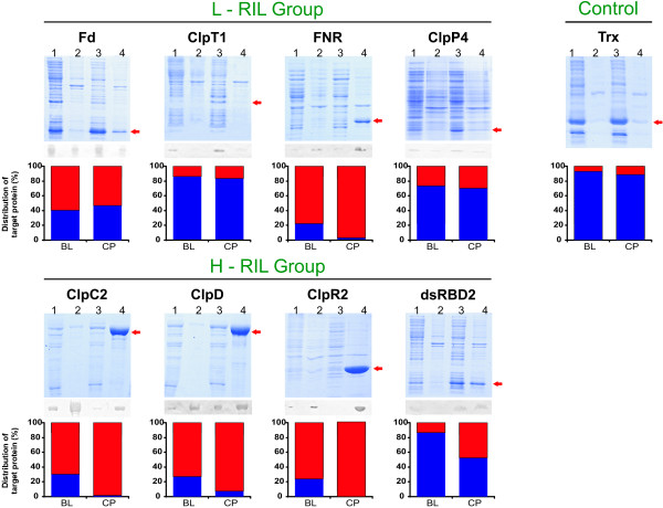
Figure 3.
Distribution of each protein in the soluble and insoluble fractions. SDS-PAGE and Western blots of soluble and insoluble fractions from induced BL (lanes 1 and 2) and CP (lane 3 and 4) cultures. Lane 1 and 3 are supernatants obtained by centrifugation of whole lysates at 10,000 g for 30 min. Lane 2 and 4 are the obtained pellets resuspended with the same volume of buffer as the supernatants. Bar plots show the percentual amount of target protein in each fraction for both strains (blue: supernatant, red: pellet). To fulfil linearity and detection limits for the inmunodetection method, a fraction of sample loaded in Coomassie Blue stained gels were loaded in Western blots as follows: FNR (lane 4), 20%; ClpC2, ClpD and ClpR2 (lane 4), 10%.

