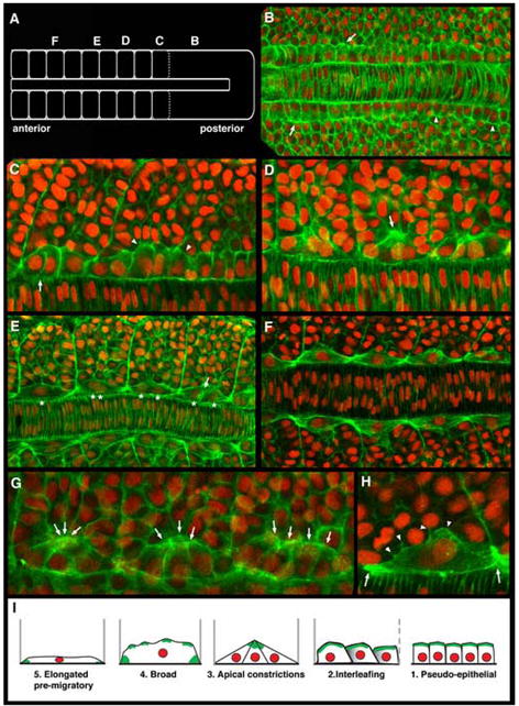
Figure 1.

Adaxial cells undergo a series of stereotypical behaviors as the zebrafish myotome matures. Confocal images of adaxial cells at the depth of the notochord within 8-10 somite stage embryos labeled with phalloidin (green) and DAPI (red). Dorsal view, anterior to the left in all panels. (A) Schematic diagram of an 8-somite stage embryo highlighting the regions where subsequent panels (B-F) are centered along the axis. (B) Adaxial cells in the posterior presomitic mesoderm region appear cuboidal (arrowheads) and become more columnar with actin-rich apices in anterior presomitic mesoderm (arrows). (C) As somite boundaries begin to form, adaxial cells begin to lose the columnar shape, displaying more irregular shapes (arrowheads) before becoming more rounded and flattened, and interleafing with one another within the dorsoventral aspect of the forming somite (arrow). (D) As the adaxial cells begin to intercalate, they transiently display actin-rich apical constrictions, with the apices of 2-4 adjacent adaxial cells apparently interacting with one another and with the more lateral cells in the central somite (arrow). (E) Following the release of the apical interactions, adaxial cells continue to broaden, as the number of adaxial cells in a given dorsal ventral section of the somite is ultimately reduced to one. Arrow shows two broad adaxial cells with interacting membranes midway between somite boundaries. Asterisks point out that as somites mature, the nuclei of broadening adaxial cells become superimposed as the adaxial cells ultimately form one dorsoventral stack of cells in the medial somite. (F) Fully intercalated adaxial cells attached to somite boundaries in the anterior somites of an 8-10 somite embryo. (G) Arrows highlight another example of the transition from early interleafing stage adaxial cells with actin-rich apices, to triangular cells displaying apical constrictions is shown across three adjacent somites. (H) Broad, intercalated adaxial cells with thin, lamellar lateral membranes appear to maintain membrane interactions with more lateral somitic cells (arrowheads), and display actin-rich, bipolar somite border attachments (arrows). (I) Schematic summary of the stereotypical behaviors displayed by adaxial cells prior to their migration.
