Fig. 6.
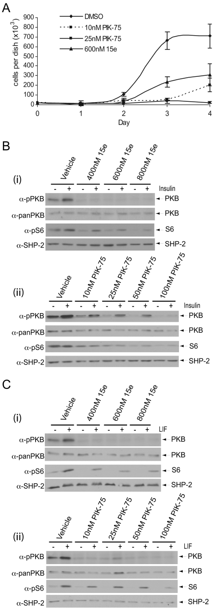
p110α appears to be coupled to proliferation, insulin signalling and LIF signalling in mESCs. (A) To assess cell proliferation, viable cell numbers were determined in triplicate at 24-hour intervals for up to 4 days. The average number of cells per dish, ± s.d., are shown for each treatment. (B,C) Following 30 minutes pre-treatment with either (i) 15e or (ii) PIK-75 at the doses indicated, mESCs were stimulated with 10 μg/ml insulin for 5 minutes (B) or 103 U/ml LIF for 10 minutes (C). Signalling downstream of PI3K was assessed by SDS-PAGE and immunoblotting with the antibodies indicated. Data, representative of three independent experiments, are shown.

