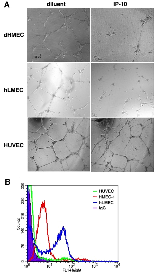
Fig. 4.
CXCR3 expression is necessary for IP-10-mediated tube dissociation. (A) dHMEC (passage 5-7), hLMEC (passage 5-7), and HUVEC (passage 3-6) were plated on GFR Matrigel in 2.0% dialyzed dFBS-EGM-2 media containing VEGF (75 ng/ml) and incubated for 24 hours to allow cord formation. The media was removed and the cells incubated in 0.5% dialyzed FBS-EGM-2 media containing VEGF (75 ng/ml) and IP-10 (600 ng/ml) then incubated for 24 hours. IP-10 was able to cause cord dissociation in dHMEC and hLMEC similar to that observed in HMEC-1. Dissociation was not observed in HUVEC. The cords were imaged with a Spot RTKE camera on an Olympus IX 70 microscope (UPlanFl 4×, 0.13). Scale bar: 150 μm. (B) It has been shown that HUVEC had a significantly lower expression of CXCR3 than microvascular endothelial cells (Salcedo et al., 2000). To verify whether the inability of IP-10 to mediate cord dissociation in HUEVCs correlated with low CXCR3 expression in these cells. HMEC-1, hLMEC and HUVEC were detached and fixed in 2% formaldehyde-Hank's balanced salt solution. The cells were incubated with anti-CXCR3 antibody conjugated with FITC and then analyzed on a BD FACSCalbur flow cytometer. The data indicate that CXCR3 expression on HUVEC (green) was just slightly above non-specific IgG (solid graph). Both HMEC-1 (red) and hLMEC (blue) had a high level of cell surface expression of CXCR3. These data suggest that the inability of IP-10 to induce HUVEC tube dissociation is due to the low surface expression of CXCR3.

