
Figure 3.
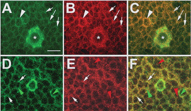
SWS and PKA-C3 colocalize in neurons. A–C, Brain whole-mounts stained with anti-PKA-C3 (green) and anti-SWS (red) show expression in most or all neurons with stronger expression of both proteins in some neurons (arrowheads), including a few large neurons, which highly express PKA-C3 (asterisks). In addition, both proteins can be colocalized to the same vesicles (arrows). D–F, In some vesicles PKA-C3 (green) colocalizes with the ER marker GRP78 (red), although both proteins can also be found separately (green arrowheads and red arrowheads). Thickness of the optical sections was 0.1 μm. Scale bar, 5 μm.