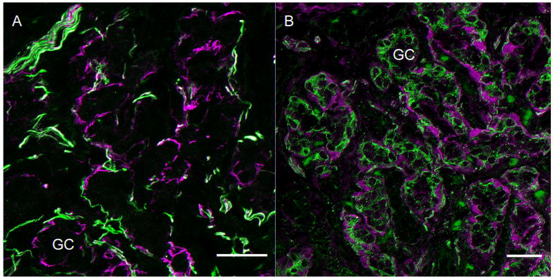
Figure 1. Markers of sensory innervation of Type 1 glomus cell clusters in the carotid body.
A. Neurofilament cocktail (anti-NF 200, 120 and 68) labels major fibers surrounding but seldom penetrating glomus cell clusters (green). Anti-GFAP (Accurate) labels cells that surround the glomus cell clusters (magenta) in close proximity to the nerve fibers. Scale bar= 25μm
B. This panel illustrates the neural innervation stained with anti-peripherin antibody. (green). The fibers surround and infiltrate the clusters of Type I glomus cells. The Type 2 cells surround the Type 1 clusters and are immunolabeled with S100 antibody (magenta, Santa Cruz). Scale bar=20μm. GC=glomus cell cluster.

