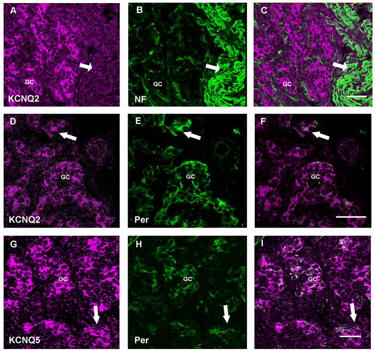
Figure 6. Expression of KCNQ channel protein in carotid body fibers.
In all panels the left image is the ion channel immunoreactivity pseudo-colored in magenta, the center image illustrates anti-NF or anti-peripherin (green). The right image is a merge of the two images. Panel A–C: Low magnification confocal z-series demonstrating the immunohistochemical localization of anti-KCNQ2 (Chemicon) with anti-NF. The arrow identifies the carotid sinus nerve. Scale = 40μm. Panel D–F: Single slice high magnification confocal image showing fine fibers with KCNQ2 (Chemicon) and peripherin (Santa Cruz) immunoreactivity in glomus cell clusters. Scale = 40μm. Panel G–I: anti-KCNQ5 overlays with anti-peripherin in fibers (arrow) and in glomus cell clusters in this confocal z-series stack. Scale = 25 μm. GC= Glomus cell clusters.

