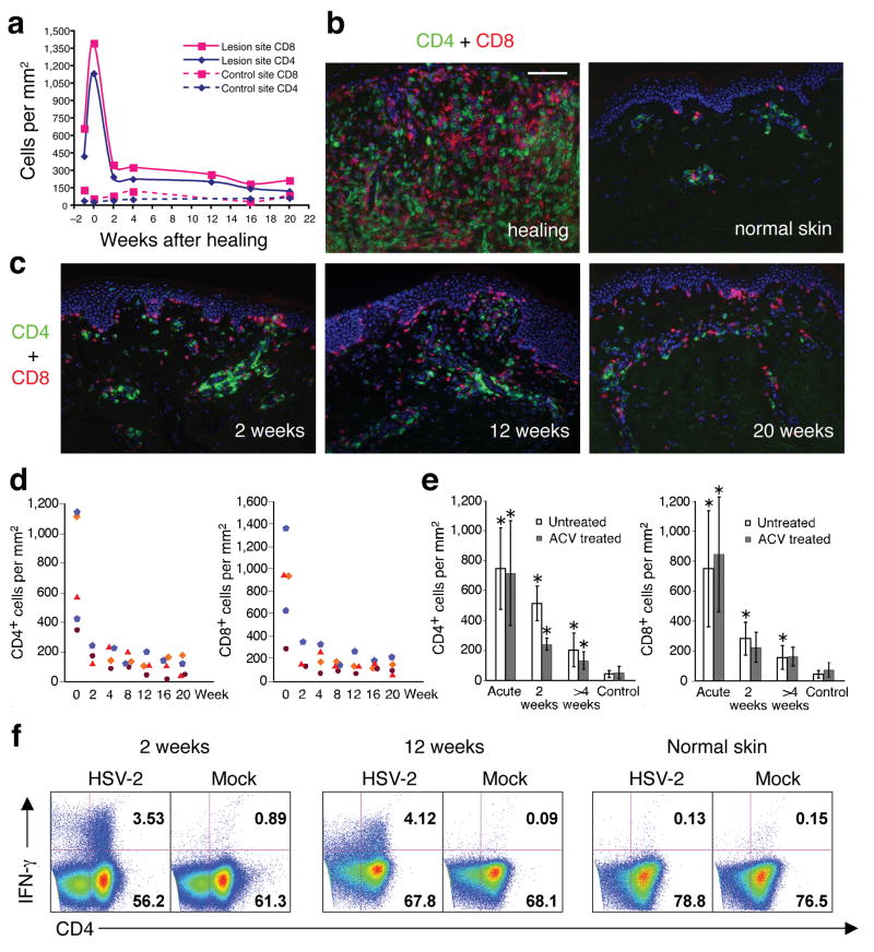
Figure 2. Persistence of CD4+ and CD8+ T cells in genital skin of subjects on daily acyclovir therapy.
(a) Quantitation of CD4+ and CD8+ cells infiltrating an HSV-2 lesion vs. uninvolved (control) genital skin. Week 0 is the time of complete lesion healing (lesion onset is labeled week -1). (b and c) Dual immunofluorescence staining of CD4+ (green) and CD8+ (red) cells. Panel b: comparison of healing stage at lesion site versus normal uninvolved genital skin. Panel c: shows CD4+ and CD8+ cells in post-healing biopsies taken from the time points indicated in panel a. (d) Quantitation of CD4+ and CD8+ cells in genital lesions of all four subjects on acyclovir therapy from acute lesion to 20 weeks post-healing. Each symbol represents a subject. (e) Comparison of CD4+ and CD8+ T cell infiltration in genital skin from untreated versus acyclovir-treated HSV recurrence. No statistically significant differences were present between acyclovir versus untreated episodes either during or after herpes reactivation (P > 0.5). The asterisk marks biopsies of statistical significance (P < 0.05) in comparison of lesion site versus control site. (f) Persistent CD4+ T cells in post-healing biopsies were HSV-2 antigen specific. After incubation with whole UV-killed HSV-2 antigen, IFN-γ expression was detected in lymphocytes collected from biopsies at 2 and 12 weeks post-HSV-2 reactivation but not in lymphocytes collected from the uninvolved genital skin (12 weeks post healing) of the subject depicted in panels a-c. Scale bar: 100 μm (B and C).

