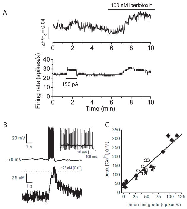
Figure 7. Block of BK channels increases intracellular calcium concentration during spontaneous firing.
A) Whole cell current clamp recordings were made from a spontaneously firing Purkinje neuron. The pipette solution included Fluo-4, a fluorescent calcium indicator. Fluorescence intensity was measured with a cooled-CCD camera. The top trace shows the increase in fluorescence intensity (ΔF) divided by the fluorescence when the cell was not firing (F0). The bottom trace is the average firing rate. At the time indicated injection of positive current (150 pA) increased both the firing rate and fluorescence intensity. At t=9 minutes, fluorescence levels in iberiotoxin were 23% greater than those in control conditions even though the average firing rate was the same. B) Tope panel. Whole cell current clamp of a Purkinje cell with a patch pipette containing Fura-4F. Injection of 200 pA of current for 1 s from a membrane potential of −70 mV evoked an average firing rate of 47 spikes/s. Bottom Panel. Simultaneous recording of intracellular free calcium concentration showed that calcium increased to about 125 nM. D) Plot of free calcium concentration versus the average firing rate. Data were collected from five cells and each symbol represents a different cell.

