Figure 1.
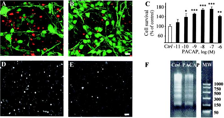
Effect of PACAP on apoptosis of cerebellar granule cells. (A and B) Typical microphotographs illustrating the effect of PACAP on survival and neurite outgrowth. Cells were cultured for 48 h in the absence (A) or presence (B) of 10−7 M PACAP. Living cells were labeled with FDA (green fluorescence), and dead cells were labeled with propidium iodide (red fluorescence). Scale bar = 10 μm. (C) Effect of graded concentrations of PACAP (10−11 to 10−6 M; 48 h) on survival of cultured cells. (D and E) Microphotographs illustrating the effect of PACAP on DNA fragmentation on cultured cells. Cells were cultured for 24 h in the absence (D) or presence (E) of 10−7 M PACAP, and DNA fragmentation was labeled by the TUNEL technique. Scale bar = 20 μm. (F) Representative gel electrophoresis illustrating the effect of PACAP on DNA laddering. MW, molecular weight markers in base pairs. *, P < 0.05; **, P < 0.01; ***, P < 0.001 vs. control (Ctrl).

