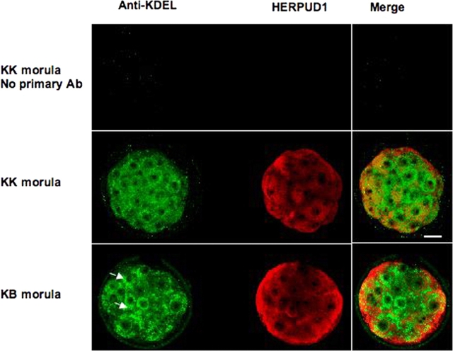
FIG. 5.
Confocal images of immunofluorescence of KK and KB morula-stage embryos. Anti-KDEL recognizes HSPA5 (detected in green), which is an integral component of ER, and HERPUD1 is linked to ER stress. Ninety-five percent of KB embryos die within 24 h of the morula stage. Arrows in KB embryo indicate areas in which ER appears to be more prominent than in KK embryos. Red fluorescence (HERPUD1) also appears more intense in KB embryo (compare merge images and see text). Ab, antibody. Bar = 20 μm.

