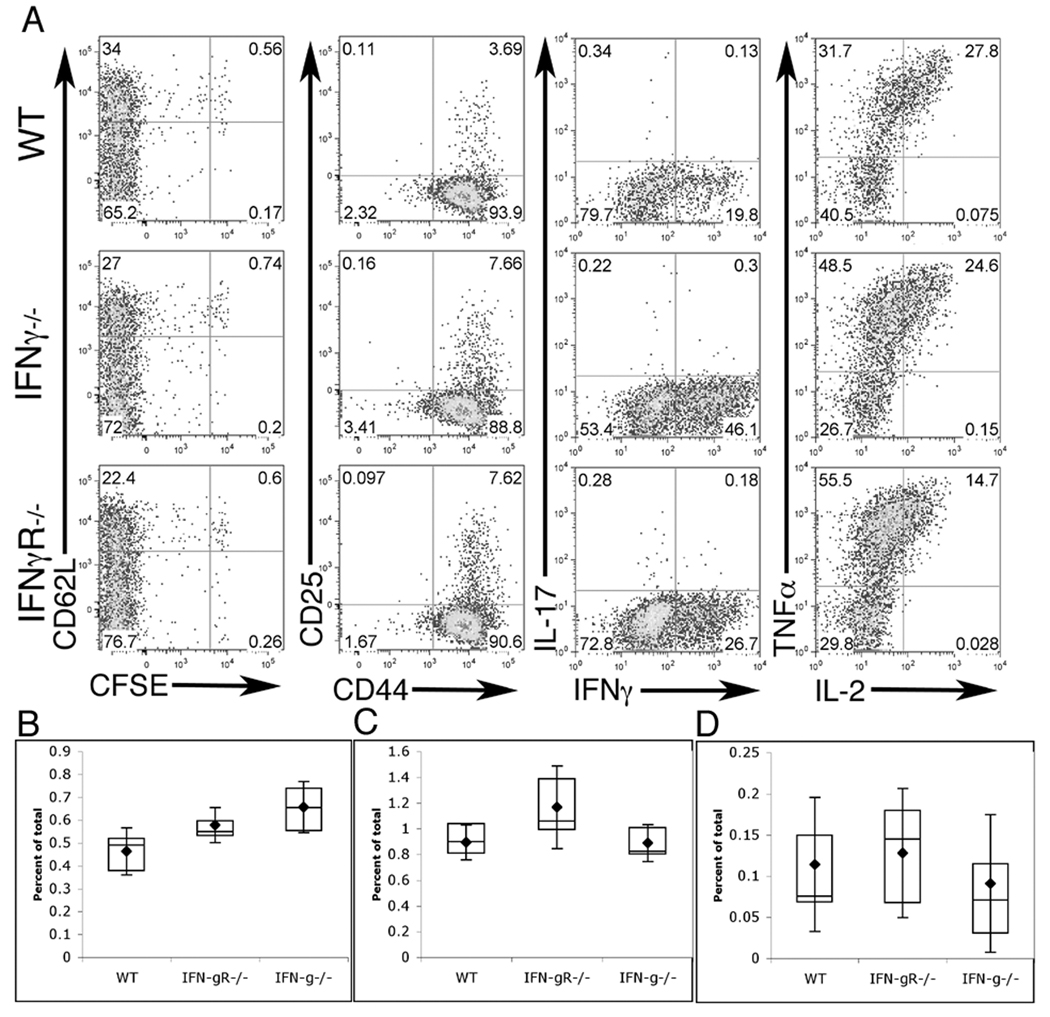
Figure 1. CD4+ T cells specific for C. trachomatis can be stimulated in IFNγ−/− or IFNγR−/− mice.
IFNγ−/−, IFNγR−/−, or WT C57BL/6 mice were injected with naïve NR1 cells and challenged the following day with C. trachomatis. On day 5 post-infection flow cytometry was used to analyze cells from the uterus, draining lymph node, and spleen (A). Cells were assessed for activation markers (left two panels) or restimulated for 5 hours with PMA/ionomoycin and assessed for intra-cellular cytokine staining (right two panels). Flow cytometry data were first gated on live, CD4+, CD90.1+ cells and are representative of three independent experiments. Percentage of total live cells was calculated for draining lymph node (B), spleen (C), and uterus (D). Statistical analysis performed via Student’s T test, * = p<0.05, ** = p<0.01.

