
Figure 1. GRP78 is differentially expressed in atheroprone areas and within atherosclerotic lesions of mice aorta.
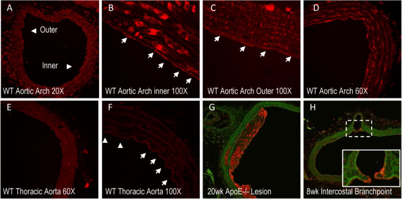
Histological sections of the aorta were stained for GRP78 (red) and matrix (green/autoflorescence) in C57BL/6 (A-F) or ApoE-/- (G-H) mice. Representative images of the aortic arch of C57BL/6 mice show the inner arch (atheroprone) and the outer arch (atheroprotective) (A-D). White arrows (100× images) indicate individual ECs. Staining was compared to protected regions of the thoracic aorta (E-F). GRP78 expressed in a 20-week lesion (G) and 8 week cross-section (H) along the descending aorta (intersecting at an intercostal branch) of an ApoE-/- mouse.