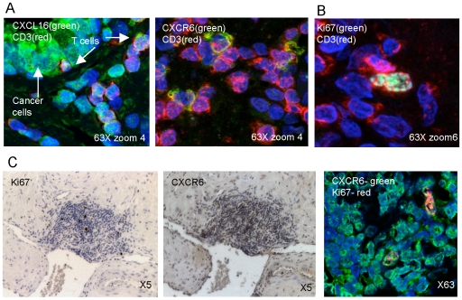
Figure 4. CXCL16 and CXCR6 are expressed by T cells in prostate tissue.
(A, B) Confocal imaging shows anti-CXCL16 (A, left panel) or anti-CXCR6 (A, right panel) or anti-Ki67 (B) as green (488 tyramide), anti-CD3 (594 tyramide) as red, and nuclear staining with Hoechst 33258 as blue. Double staining in yellow shows co-localization. In (A), arrows point to cancer cells and T cells. Panels are representative of more than 15 cases for each double-stain. (C) Staining for Ki67 and CXCR6 in a region of prostatitis using DAB (brown, two left panels) or immunofluorescence (right panel) for detection. Confocal imaging shows anti-CXCR6 as green (488 tyramide) and anti-Ki67 as red (594 tyramide), and nuclear staining with Hoechst 33258 as blue. Panel is representative of more than 15 cases.

