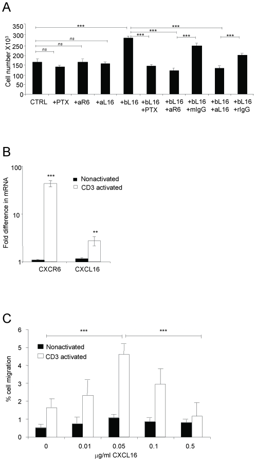
Figure 6. CXCL16 can mediate the proliferation and migration of CD3-activated primary CD4 T cells.
(A) CD4+ T cells purified from elutriated lymphocytes from healthy donors were stimulated for 3 days with plate-bound anti-CD3 (OKT3, 10 µg/ml) with or without 5 µg/ml plate-bound CXCL16 (bL16). Treatments of anti-CD3-activated cells with PTX, anti-CXCR6 (aR6) and anti-CXCL16 (aL16) without bL16 were done as controls. Mouse IgG (mIgG) and rat IgG (rIgG) were used as controls for anti-CXCR6 antibody and anti-CXCL16 antibody, respectively. Bars show means±SEM from one representative experiment out of five, using five donors. Anti-CXCL16 was used in a total of four, and anti-CXCR6 in two experiments. ns, not significant and ***, p<0.001. (B) CXCR6 and CXCL16 mRNAs were measured by real-time RT-PCR in CD3-activated and non-activated CD4+ T cells after 3 days. Values were normalized by setting the non-activated sample with lowest expression equal to 1. Bars show means±SEM combined from duplicate wells from seven different donors. **, p<0.01 and ***, p<0.001 vs. non-activated cells. (C) Anti-CD3-activated or nonactivated CD4+ T cells were analyzed for migration to CXCL16, expressed as percentages of input cells migrating. Each bar represents the mean±SEM obtained from triplicate wells from a total of three experiments performed. ***, p<0.001 on cross bars are indicated for comparisons between activated cells - 0 vs. 50 ng/ml CXCL16 and 50 ng/ml vs. 500 ng/ml CXCL16.

